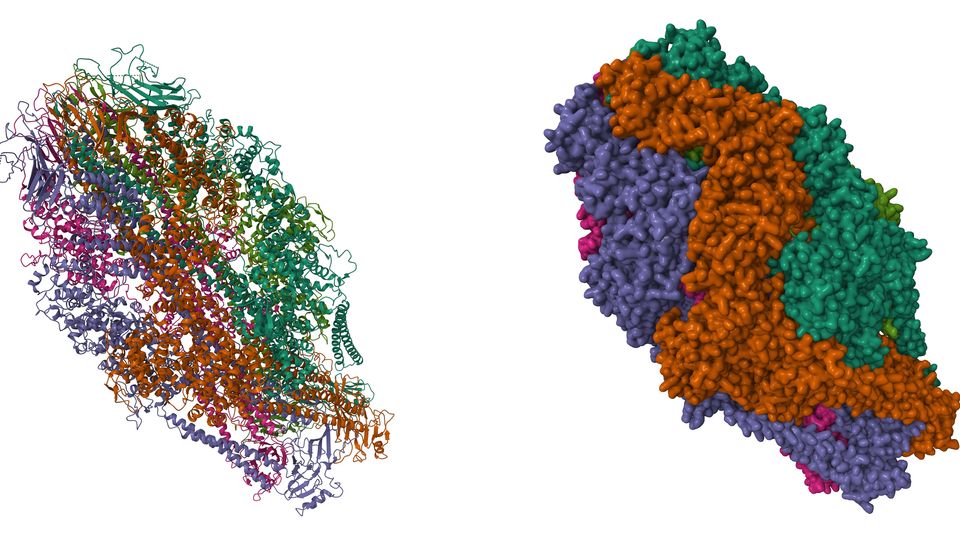
Microscopic imaging has revolutionized the way we perceive the world around us, delving into the intricacies of objects at a minuscule scale. The advent of three-dimensional (3D) imaging techniques has further empowered researchers and enthusiasts to explore the depth and structure of microscopic specimens. In this article, we will unravel the capabilities of two prominent microscopes that have the potential to generate stunning three-dimensional images.

Credit: www.technologynetworks.com
Confocal Microscope: Peering into the Depths with Precision
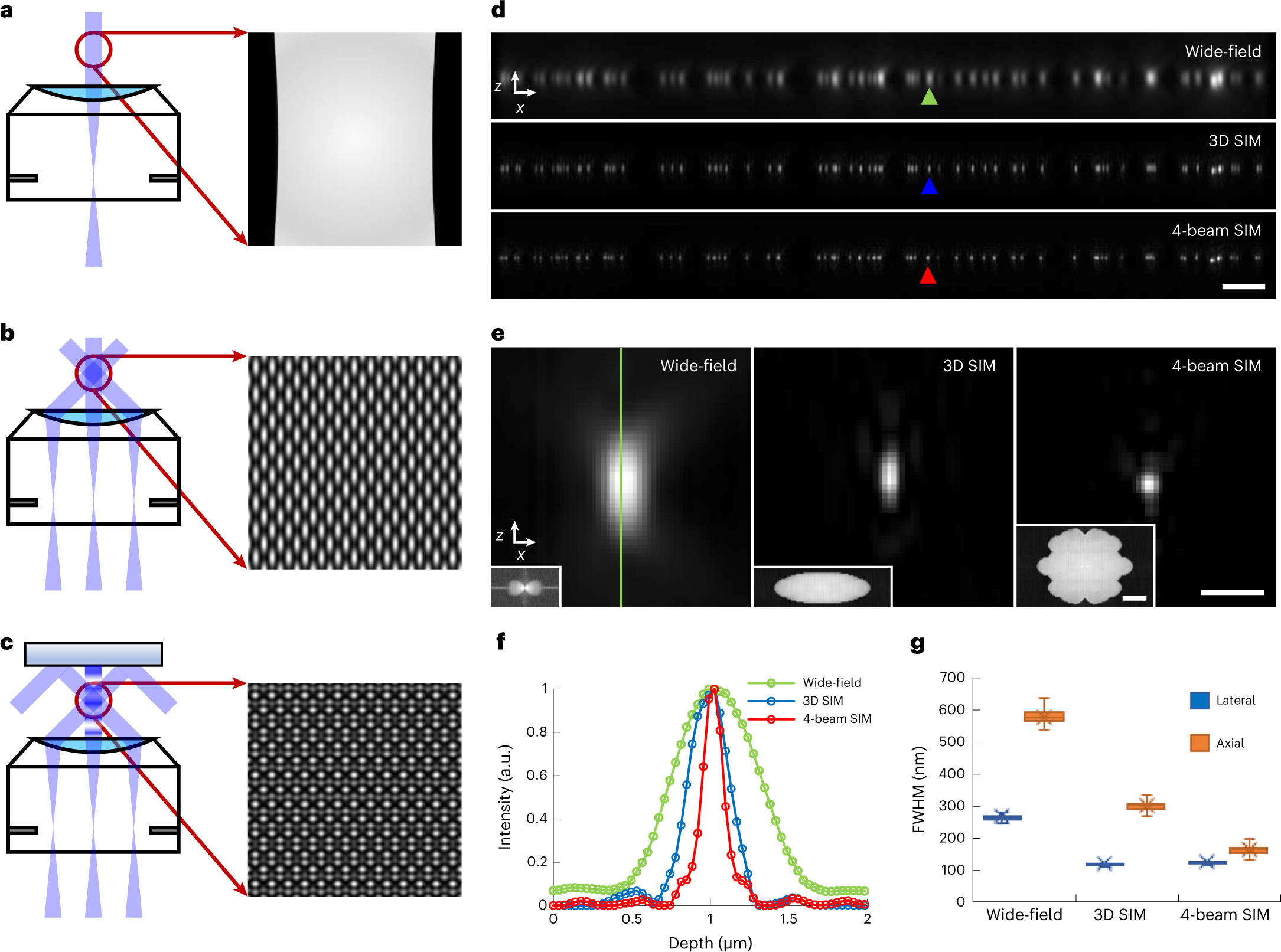
The confocal microscope is a powerful tool that has contributed significantly to three-dimensional imaging in biological and material science research. The key attribute of a confocal microscope lies in its ability to capture images from different focal planes, subsequently amalgamating them to reconstruct a detailed 3D representation of the specimen.
This meticulous process involves the use of a pinhole to selectively allow light from the focal point to pass through while blocking out-of-focus light, resulting in exceptionally sharp, high-contrast images. Through this mechanism, the confocal microscope offers unprecedented precision and depth of field, enabling researchers to visualize complex biological structures and surface morphology with remarkable clarity.
| Advantages of Confocal Microscope for 3D Imaging |
|---|
| High precision and clarity in capturing 3D structures |
| Ability to eliminate out-of-focus light, enhancing image quality |
| Accurate visualization of intricate biological and material samples |
Atomic Force Microscope (AFM): Unraveling Nanoscale 3D Landscapes
Delving into the realm of nanoscale exploration, the Atomic Force Microscope (AFM) emerges as a pivotal instrument for capturing three-dimensional images with remarkable precision. Operating on the principle of scanning a sharp probe across the specimen surface, the AFM detects interactions between the probe and the surface, translating these interactions into a three-dimensional topographic map.
One of the standout features of AFM is its versatility, enabling researchers to explore diverse samples including biological molecules, polymers, and semiconductor structures at the nanoscale. This capability makes AFM an indispensable tool for unveiling the intricate 3D landscapes of nanomaterials and biological entities.
| Advantages of Atomic Force Microscope for 3D Imaging |
|---|
| High-resolution 3D imaging at the nanoscale level |
| Versatile applicability across various sample types |
| Visualization of nanoscale surface morphology and properties |

Credit: www.nature.com
Integrating Three-Dimensional Imaging into Diverse Disciplines
The ability to generate three-dimensional images holds immense potential across a wide spectrum of scientific and industrial domains. In the field of life sciences, 3D imaging techniques facilitated by confocal microscopes have enabled detailed investigations into cellular structures, tissue architecture, and developmental processes, thereby enhancing our understanding of biological phenomena.
On the industrial front, the utilization of AFM for 3D imaging has proven crucial in nanotechnology, materials science, and semiconductor research, empowering engineers and scientists to analyze surface roughness, mechanical properties, and nanoscale features of diverse materials and devices.
Furthermore, the application of three-dimensional imaging extends to the realms of archaeology, art conservation, and forensics, where the visualization of intricate surface details and structural nuances plays a pivotal role in deciphering historical artifacts, preserving cultural heritage, and aiding in criminal investigations.
The Future of Three-Dimensional Microscopic Imaging
As technological advancements continue to unfold, the landscape of three-dimensional microscopic imaging is poised for further evolution. Innovations in microscopy techniques, computational imaging, and artificial intelligence-based image analysis hold the promise of enhancing the resolution, speed, and interpretability of 3D imaging, opening new frontiers for research and exploration at the microscopic level.
With the convergence of multidisciplinary expertise and cutting-edge technologies, the potential applications of three-dimensional imaging in fields such as medicine, materials engineering, environmental science, and beyond are bound to expand, ultimately offering profound insights into the intricate realms of the microscopic world.
Embracing a future where three-dimensional imaging becomes more accessible, versatile, and insightful, the quest to unravel the mysteries of the microscopic universe continues, empowering researchers, educators, and innovators to embark on transformative journeys driven by the power of microscopic visualization.
Frequently Asked Questions For Which Two Microscopes Generate Three Dimensional Images: Advanced Technology Revealed
What Are The Two Microscopes Used For Generating Three-dimensional Images?
Two microscopes commonly used for generating three-dimensional images are the confocal microscope and the electron microscope.
How Does A Confocal Microscope Generate Three-dimensional Images?
A confocal microscope generates three-dimensional images by capturing optical sections at different depths within the specimen and then reconstructing them using computer software.
What Are The Advantages Of Using A Confocal Microscope For Three-dimensional Imaging?
Using a confocal microscope for three-dimensional imaging provides advantages such as better resolution, elimination of out-of-focus blur, and the ability to observe living and fixed specimens.
How Does An Electron Microscope Generate Three-dimensional Images?
An electron microscope generates three-dimensional images by using a beam of electrons instead of light to form an image, with the help of computer software for reconstruction.

